What Is a Chest Tube?
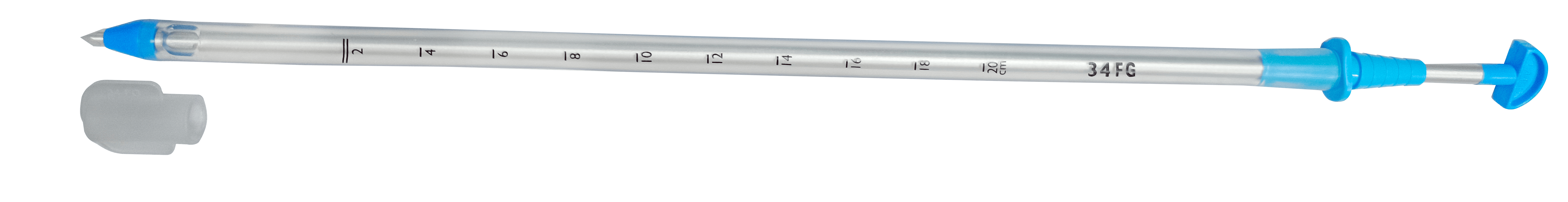
A chest tube is a plastic tube that is utilized to remove liquid from the chest. Liquid (for case blood or discharge) that collects in the space between the lungs and chest wall (which is also called the pleural space) can cause the lung to collapse. Chest tubes come in different shapes and sizes. Depending upon its application, they have different sizes based on their application.
Chest tubes helps to collect liquid and permit liquid tpo to drain from the chest. These frameworks can be manual or it can have some mechanism to flow out the liquid using some suction pressure applied to it.
Pre-Procedure Precautions Ensuring Comfortable and Safe Placement of Tube
- The patient will lie on their back or be subtly propped up before a tube placement procedure
- The area where the nurse will operate will have to be extremely clean and free of germs, usually on one side of the chest. We make sure that the procedure is safe in an already sterile area.
- Before proceeding, Need to give some numbing medicine right where we’re going to put the tube.This will ensure that you won’t be in any pain when it is being inserted so it’s as pain-free as possible.
Steps for Tube Placement:
- A small incision is made in the skin when the tube is inserted, which is usually on the chest, opposite the armpit, and between the ribs.
Why This Spot? - It’s to ensure that to select a very specific area so nothing bad, such as blood vessels or nerves, ensuring the procedure is safe and effective.
Accessing the Pleural Space: What’s Next?
In the procedure of tube placement, we gain entry to the pleural space that surrounds the lungs. And here’s how it is done.
Creating a Passage:
Now slowly advance across the layers of skin, tissue, and muscle,since this is the tme to make our way toward the region that surrounds the lungs.
Being Cautious:
At this point, care should be taken as there is a possibility of injury to other organs located besides the pathway.
Placement of a Chest Tube: Step-by-Step Explanation
1. Insertion of the Chest Tube
After the access into the thoracic cavity, use a trochar or a blunt-tipped dilator to create a larger hole. Then insert a pliable tube into the newly created hole. This tube is perforated along its sides by tiny holes, which allow excess fluid or air to leak out.
Now connect this tube with a drainage system, so that the fluid or air is evacuated out smoothly in the body, and the patient feels better soon.
2. Verification and Fixation
After inserting the chest tube, check if it is in the right position or not. Most of the times it got fixes in proper position and sometime an X-ray is taken for verification purposes.
We secure the tube in place either by sutures, special tape, or a specific device that will keep it from moving, after having ensured it has reached the appropriate site.
3. Monitoring and Post-Procedure Care
Monitor the patient’s respiratory sounds and vital signs, including heart rate and blood pressure, also monitor the amount of fluid draining through the tube.
Sometime immediate measures is taken if there is a problem such as lung collapse, hemorrhage, or infection. It is been checked regularly to make sure that the chest tube is really working well.
Understanding the Risks
Insertion of a chest tube is normally safe. But, as with every procedure, there are some risks involved:
The risk includes that of bleeding at the insertion site or in the chest; this is much worse in those patients with haematologic disorders or who are treated with blood thinners.
- Infection: A complication which can arise due to an infection as a result of the placement of the tube, especially from the entry of bacteria during the placement of the tube, such as pneumonia or infection occurring at the insertion site.
- Pneumothorax: Sometimes, placing a chest tube can inadvertently cause a pneumothorax wherein air gets into the space surrounding the lungs and may collapse a lung. Though this is quite rare. Keeping a close Eye in this process is important.
- Subcutaneous Emphysema: Sometimes, air may leak into the tissue immediately below the skin and causes swelling and a crunching feel when touched.
- Organ Injury: There is a potential for accidental injury to nearby organs, such as the lungs or liver, when this is performed.
- Pain and Discomfort: Most patients experience some degree of pain or soreness at the site where the tube is inserted after the procedure.
- Malposition: Sometimes, the chest tube is not placed well. This can lead to poor drainage of fluid or air.
- Re-expansion Pulmonary Edema: There may be significant fluid accumulation in the lungs after re-expansion of a collapsed lung.
Differences Between Chest Drainage Catheters With and Without Trocar:
1) Chest Drainage Catheters With Trocar-
- Design: It is attached with sharp pointed tool called trocar, which will perforate the chest wall for placing the catheter inside the patient.
- Insertion Technique: The trocar will directly penetrate the chest wall and inside the pleural space an opening will be formed. The catheter will then be inserted with much ease.
Advantages:
- Speed: For those who have trocar, the introduction is quicker since the trocar will puncture the chest wall directly.
- Ease: In emergency conditions, it will be easier and faster to relieve the pressure
Disadvantages
- Risk of Injury: The sharp point of the trocar has more chances of reaching the internal structures accidentally like lung and blood vessels.
- Poor Control: A catheter with less control about the depth as well as the angle of insertion than all other methods.
2) Chest Drainage Catheters Without Trocar
- Design: No trocar is present in these catheters. These catheters are placed using a guidewire (Seldinger technique) and by blunt dissection.
- Insertion Technique: Guidewire Method or Seldinger Technique: By the guidewire, puncturing initially through the needle is done into pleural space. A thin guidewire is passed through the hole of the needle. The needle is withdrawn and over the wire catheter passes.
- Blunt Dissection: An incision of a few millimeters is created and tissues are gently separated for passage of the catheter.
Advantages:
- I.Safety: These techniques prevent damage to major structures by accident as this technique provides more control to the operator during the insertion process.
- Accuracy: These techniques also provide a more precise positioning of the catheter.
Disadvantages:
- I.Time: This procedure will take more time
- II.Technical Complexity: The Seldinger technique demands more technical knowledge and technical capability.
Clinical Considerations
Emergency Conditions: Doctors may use only the trocar catheter to aspirate pressure as fast as possible in conditions of emergency.
Elective Conditions: Usually, doctors do not use techniques involving trocar in the case of non-emergency situations for they are safer and much more precise.
Patient Anatomy: Based on the patient anatomy, and also on the character of fluid or air which is to be aspirated, the selection of the method usually depends on the specifics of the medical condition.
Advantages of a Chest Tube
Chest tube offer several benefits to patients if the condition being treated so requires. Among the most important are these:
- 1. Improved lung function: A collapsed lung or fluid that may have been exerting some compressive force on the lung is sucked out by the insertion of a chest tube, allowing the lung to expand and thus ease breathing.
- Prevention of Complications: Pneumothorax would be a collapsed lung or hemothorax-blood in the chest if it were not taken seriously, then serious complications can come to occur. The chest tube allows appropriate expansion to be given by draining air or fluid from the chest.
- Symptoms and signs relieved: chest tubes relieve symptoms due to pleural effusion-in this case, chest pain, difficulty breathing, and cough-and therefore provide a comfort for quality of life for a patient.
- Making other treatments easier: chest tubes are often useful in assisting other treatments such as pleurodesis (to prevent secondary fluid accumulation) or thoracic surgery, creating an environment more amenable to those treatments.